| |
||||||||||
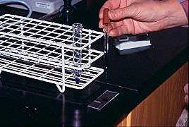
| Faecal sedimentation: Procedure step 10 |
Purpose |
|||
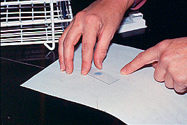
| Transfer a small drop of the stained sediment to a microscope slide using a pipette. Cover droplet with a coverslip.
Examine under a microscope at 10 x 10 magnification. Repeat until all the sediment has been examined. Alternatively, pour the whole amount into a Petri dish and examine methodically under a stereo-microscope.
Warning!!! Nematode eggs may be present in the faecal
sample but the recovery with this technique is very low. Nematode
eggs should be examined using flotation
techniques or the MacMaster
technique.
|
|||