
Page 8 - Clinical Connections - Spring 2024
P. 8
RVC RESEARCH STUDY VETERINARY SERVICES RVC.AC.UK
Orthopaedics
LATEST IN GAIT ASSESSMENT WITH THE
ORTHOPAEDIC SERVICE
Richard Meeson, Professor of Orthopaedics and Head of the Orthopaedic Service
ecently, thanks to the kind generosity and for monitoring response to treatment.
of the Animal Care Trust (ACT), the Over the last 10 years, the Queen
R RVC’s registered charity, a grant Mother Hospital for Animals has used a
was awarded to upgrade the now out of flat pressure-mat, which measures the
date pressure mat, to allow the team to pressure from each paw as a dog walks
expand their lameness diagnosis and across it. Technology and the computer’s
clinical monitoring through a state-of-the-art software have improved greatly since then
instrumented dog treadmill. and a further development was to combine
The treadmill is similar to that used by this with a treadmill. For the first time, it is
humans in gyms, however this one blends possible to have a dog walking consistently
a state-of- the-art matrix of tiny pressure without ‘running out of runway’ and to
sensors. A computer then matches the evaluate them at faster speeds.
pressure sensor activation to the correct For dogs that are not very mobile, it will
foot, from the synchronised cameras filming even give a standing evaluation of how
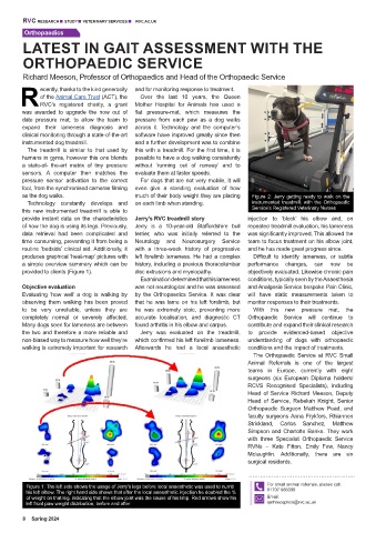
as the dog walks. much of their body weight they are placing Figure 2: Jerry getting ready to walk on the
Technology constantly develops and on each limb when standing. instrumented treadmill, with the Orthopaedic
this new instrumented treadmill is able to Service’s Registered Veterinary Nurses
provide instant data on the characteristics Jerry’s RVC treadmill story injection to ‘block’ his elbow and, on
of how the dog is using its legs. Previously, Jerry is a 10-year-old Staffordshire bull repeated treadmill evaluation, his lameness
data retrieval had been complicated and terrier, who was initially referred to the was significantly improved. This allowed the
time consuming, preventing it from being a Neurology and Neurosurgery Service team to focus treatment on his elbow joint,
routine ‘bedside’ clinical aid. Additionally, it with a three-week history of progressive and he has made great progress since.
produces graphical ‘heat-map’ pictures with left forelimb lameness. He had a complex Difficult to identify lameness, or subtle
a simple overview summary which can be history, including a previous thoracolumbar performance changes, can now be
provided to clients (Figure 1). disc extrusions and myelopathy. objectively evaluated. Likewise chronic pain
Examination determined that his lameness conditions, typically seen by the Anaesthesia
Objective evaluation was not neurological and he was assessed and Analgesia Service bespoke Pain Clinic,
Evaluating ‘how well’ a dog is walking by by the Orthopaedics Service. It was clear will have static measurements taken to
observing them walking has been proved that he was lame on his left forelimb, but monitor responses to their treatments.
to be very unreliable, unless they are he was extremely stoic, preventing more With this new pressure mat, the
completely normal or severely affected. accurate localisation, and diagnostic CT Orthopaedic Service will continue to
Many dogs seen for lameness are between found arthritis in his elbow and carpus. contribute and expand their clinical research
the two and therefore a more reliable and Jerry was evaluated on the treadmill, to provide evidenced-based objective
non-biased way to measure how well they’re which confirmed his left forelimb lameness. understanding of dogs with orthopaedic
walking is extremely important for research Afterwards he had a local anaesthetic conditions and the impact of treatments.
The Orthopaedic Service at RVC Small
Animal Referrals is one of the largest
teams in Europe, currently with eight
surgeons (six European Diploma holders/
RCVS Recognised Specialists), including
Head of Service Richard Meeson, Deputy
Head of Service, Rebekah Knight, Senior
Orthopaedic Surgeon Matthew Pead, and
faculty surgeons Anna Frykfors, Rhiannon
Strickland, Carlos Sanchez, Matthew
Simpson and Charlotte Banks. They work
with three Specialist Orthopaedic Service
RVNs – Kate Fitton, Emily Few, Nancy
Mclaughlin. Additionally, there are six
surgical residents.
Figure 1: The left side shows the usage of Jerry's legs before local anaesthetic was used to numb For small animal referrals, please call:
his left elbow. The right hand side shows that after the local anaesthetic injection he doubled the % 01707 666399
of weight on that leg, indicating that the elbow joint was the cause of his limp. Red arrows show his Email:
left front paw weight distribution, before and after qmhreception@rvc.ac.uk
8 Spring 2024