
Page 6 - Clinical Connections - Summer 2023
P. 6
RVC RESEARCH STUDY VETERINARY SERVICES RVC.AC.UK
Advanced imaging
SPOTLIGHT ON IMAGING-GUIDED
PROCEDURES
Francisco Llabres-Diaz, Head of the Small Animal Diagnostic Imaging Service and Senior Lecturer in
Veterinary Diagnostic Imaging
iagnostic imaging has developed at sample, not only because of their small size, they cannot be removed under ultrasound
an amazing pace in the last 20 years. but also sometimes due to their location in guidance.
DWe can now obtain hugely detailed the body or the fact that they are very close Although the colleagues in the
images of the body that continuously help the to vital structures like large blood vessels. anaesthesia team perform locoregional
Queen Mother Hospital for Animals (QMHA) Samples from orbital abscesses causing anaesthesia, rather than the imaging team,
offer better patient care. The main areas of exophthalmos are another area where it is worth mentioning that ultrasound is
change in this period include quick in-house imaging can help not only diagnose but also used in many of those cases to guide the
access to advanced imaging modalities (CT treat. procedure, demonstrating the wide uses of
and MRI) and the quality of the images that imaging throughout the hospital.
are obtained (this applies to all modalities, Specialist training Ultrasound is, however, not the only
with the arrival of digital radiography and the We can teach residents how to safely guiding imaging modality at the QMHA.
software and hardware improvements seen perform these procedures after a relatively Fluoroscopy is routinely used by our
in ultrasound). short period of time. From July, the imaging colleagues in the anaesthesia team to
In addition, the Diagnostic Imaging Service team will be training five residents to become perform epidural injections in the pain clinic
at the QMHA has extensive experience in specialists in imaging. We are a large (and and the radiology team has been using
helping patients through imaging-guided successful) training centre where many CT to guide the sampling of deeper or
procedures. Within those, ultrasound is veterinary surgeons interested in imaging more challenging lesions that may not be
by far the modality most frequently used apply for further training. immediately visible with ultrasound.
and, within ultrasound-guided procedures, We can also use ultrasound to guide A sad case where CT-guided sampling
ultrasound-guided fine needle aspirates are the placement of needles that help the soft helped the owners receive confirmation that
the most commonly performed. Examples tissue surgeons find smaller structures that their beloved dog suffered from a tumour
would include obtaining cells from the liver need to be removed surgically. Special affecting the base of the skull demonstrated
and the spleen or masses. localisation needles originally designed, this well. We would not have been able to
The team is also experienced in targeting for instance, for locating breast lesions in sample the lesion with ultrasound.
far smaller and challenging structures. It human medicine, are used. Once deployed, Imaging-guided sampling and therapy
is not uncommon for colleagues in other they leave a guiding wire within the patient. is a growing field. Interventional radiology
departments to ask us to perform fine- The surgeon can then follow that wire to the is becoming more widespread in multiple
needle aspirates of small lymph nodes in the target lesion, with a significant decrease in veterinary centres. The imaging team is not
region of 2 to 3 mm in width. surgical time. Foreign bodies can also be directly involved in this field at the QMHA, but
These lymph nodes can be difficult to marked for removal in a similar fashion, if fluoroscopy is heavily used in interventional
procedures.
Examples of areas where the imaging
team could expand its role include the use
of MRI. Here, the use of MRI-safe needles,
together with the superb contrast resolution
of MRI, can help in cases where MRI has
been the modality of choice to investigate
the clinical signs. Furthermore, imaging
could become involved in the use of ablation
techniques to treat lesions.
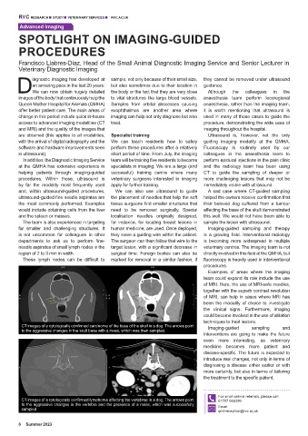
CT images of a cytologically confirmed carcinoma of the base of the skull in a dog. The arrows point Imaging-guided sampling and
to the aggressive changes in the skull base with a mass, which was then sampled.
interventions are going to make the future
even more interesting, as veterinary
medicine becomes more patient and
disease-specific. The future is expected to
introduce real changes, not only in terms of
diagnosing a disease either earlier or with
more certainty, but also in terms of tailoring
the treatment to the specific patient.
For small animal referrals, please call:
CT images of a cytologically confirmed lymphoma affecting the vertebrae in a dog. The arrows point 01707 666399
to the aggressive changes in the vertebra and the presence of a mass, which was successfully
sampled Email:
qmhreception@rvc.ac.uk
6 Summer 2023