
Page 4 - Clinical Connections - Autumn 2022
P. 4
RVC RESEARCH STUDY VETERINARY SERVICES RVC.AC.UK
Equine
PAIN(T) IN THE NECK – CT MYELOGRAPHY
OF THE CERVICAL SPINE
Maty Looijen, Equine Resident
linical problems associated with cerebrospinal fluid protects the spinal cord). compression within the neck.
the cervical spine in horses are It is historically used to identify spinal cord There are a variety reasons for
C common. They involve a wide variety compression causing ataxia. However, the compression, but cervical vertebral
of symptoms, including neurological deficits myelographic contrast column abnormalities malformation is one of the most common
(e.g. ataxia, dysmetria, paresis), neck associated with the abnormality of the causes of ataxia in sport horses. It is
pain and stiffness, and gait abnormalities. vertebral canal can only be accessed thought to be a developmental abnormality
Neurological abnormalities most commonly in a single dorsoventral orientation on caused by genetic predispositions and
result from spinal cord abnormalities (e.g. radiographs. environmental influences, such as diet,
infection/inflammatory conditions, neoplasia growth rate, workload and trauma
or compression). The pathophysiology of the disease
Investigation of horses with neck problems involves spinal cord compression due to
can involve a neurological and/or lameness malformation and/or malarticulation of the
examination, blood tests, a cerebrospinal vertebrae, static or dynamic instability of
fluid tap or an EMG (electromyography the vertebral canal, soft tissue or bony
– electronic muscle stimulant test). changes of the cervical vertebral bodies,
Radiography and ultrasonography may be their articulations and associated soft tissue
helpful tools in investigating the cervical Figure 2: Transverse CT image at the level of structures.
C4-C5, on the left without myelogram and on
spine, however they lack sensitivity and the right with myelogram
specificity.
Radiographic examinations are Advanced imaging
commonly performed for the evaluation of MRI and CT are much better modalities
potential bone abnormalities and alignment to evaluate and further identify the cause
of the vertebrae. If abnormal findings are of spinal cord compression, due to their
seen, a further challenge is to distinguish 3D compilation of the anatomy. MRI of
their clinical significance. This is similar for the cervical spine in horses is currently
ultrasonographic examinations, which are not available, due to the limited size of the Figure 3: Transverse CT image at the level
commonly performed to further investigate magnet. CT, however, has become popular of C6-C7 showing moderate reduction of the
dorsal contrast column indicating focal spinal
the soft tissues associated with the cervical over the last decade. Additionally, the spinal cord compression
spine. The neck has a complex anatomy, cord can be highlighted by CT myelography,
and radiography or ultrasonography are which now can be investigated in a Furthermore, we have to distinguish
not able to visualise details of the deeper lateromedial and craniocaudal direction abnormalities which originate from either
structures (e.g. the spinal cord). Both as well as dorsoventral. By visualising degenerative or acquired lesions, such as
modalities therefore often lack the possibility lesions accurately, we can improve our osteoarthritis, fractures, (sub)luxations,
to establish a final diagnosis. understanding of cervical spine pathologies, intervertebral disc disease or neoplasias.
treat lesions more precisely and develop Osteoarthritis of the articular process joints
new treatments. does not necessarily implement compression
A CT myelography of the neck at the RVC on the spinal cord, this depends on their
is performed under general anaesthesia size and shape in relation the position of
to avoid side effects and improve image the spinal cord in the vertebral canal. Lastly,
quality. Contrast medium is injected into the soft tissue abnormalities commonly found in
subarachnoid space, at the atlanto-occipital relation to spinal cord compression involve
articulation (figure 1). synovial cysts, epidural haematomata or
It diffuses in the vertebral canal caudally ligamentum flavum thickening – all which
and thereby outlines the spinal cord (figure we now diagnose with the help of CT
2). Interpretation is done by our radiologists myelography.
via subjective evaluation and objective
measurements, indicating compression
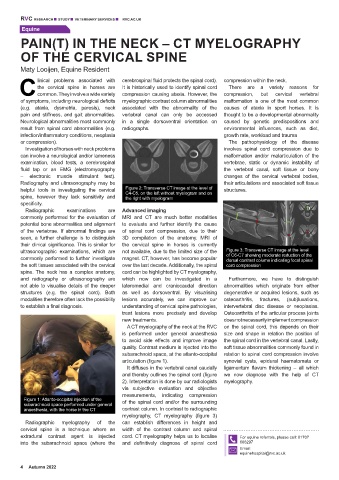
Figure 1: Atlanto-occipital injection of the of the spinal cord and/or the surrounding
subarachnoid space performed under general
anaesthesia, with the horse in the CT contrast column. In contrast to radiographic
myelography, CT myelography (figure 3)
Radiographic myelography of the can establish differences in height and
cervical spine is a technique where an width of the contrast column and spinal
extradural contrast agent is injected cord. CT myelography helps us to localise For equine referrals, please call: 01707
into the subarachnoid space (where the and definitively diagnose of spinal cord 666297
Email:
equinehospital@rvc.ac.uk
4 Autumn 2022