24 hour contact: 01707 666297
Computed Tomography (CT)
Computed tomography is a technique that allows us to produce 3D x-ray images of your horse's head, neck and legs.
The RVC Equine Referral Hospital was the first equine hospital in the UK to install a CT scanner for horses, in 2003. Our equine specialists have scanned hundreds of horses since, thus accumulating a huge amount of expertise in this advanced diagnostic imaging method. Our equine vet team are experts in acquiring the images smoothly and interpreting them accurately in an efficient manner.
The Qalibra CT scanner system we use allows the imaging of the head and upper part of the neck in the standing horse. This is ideal for diagnosing teeth and sinus problems. The scanner enables further evaluation of horses who sustained trauma to their head or show signs of headshaking. It is also a very helpful technique for surgery planning and evaluation of treatment outcomes.
Due to the large 90cm bore of the scanner, under general anaesthesia, there are further regions that can now also be imaged. This includes:
- entire neck
- pelvis
- sacroiliac joints
- proximal limbs
- cervicothoracic junction (the segment connecting the neck with the upper back). This is especially helpful for horses showing neck pain, unexplained forelimb lameness and/or ataxia (neurological problems) and in which conventional x-rays and ultrasounds were previously inconclusive or normal
- To book, call us on 01707 666297 or submit your (non-urgent) referral request online
How does an equine CT scanner work?
CT uses a rotating x-ray machine to produce a stack of slice-like images of the body.
This has the advantage that all structures are clearly displayed in cross-section and there is no superimposition of structures that hinder interpretation of the images.
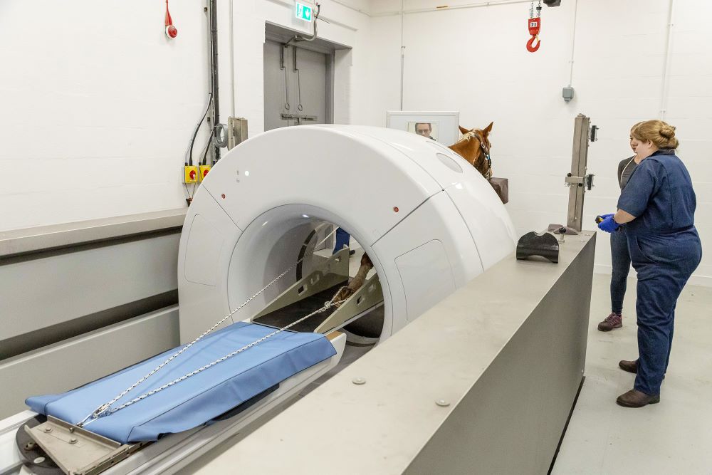
- When images are being acquired, the patient is slowly moved through the circular tunnel of the CT scanner.
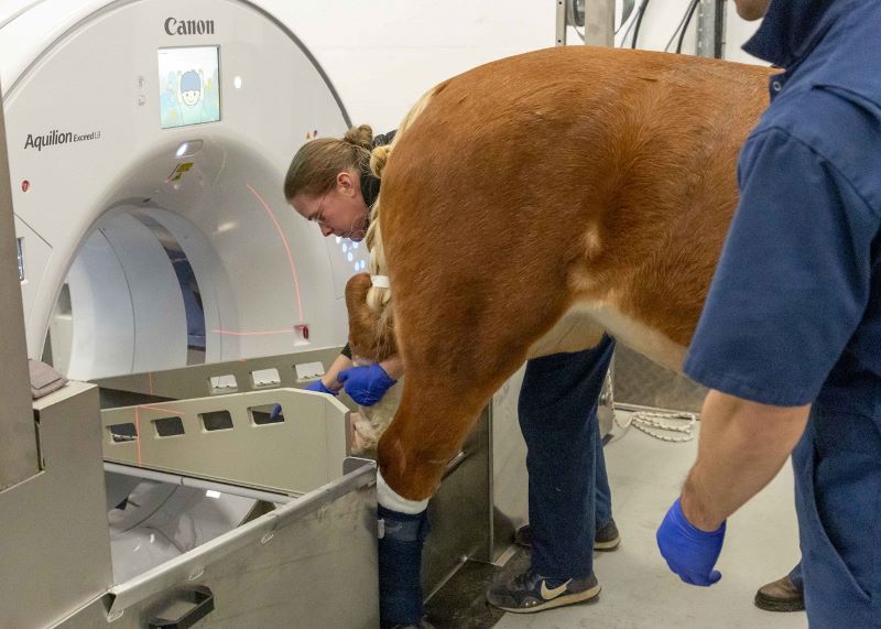
- The head and upper part of the neck can be scanned in the standing, sedated horse, for this we stand the horse on an air hover platform.
- Since the scanner is noisy we usually put cotton wool in the horse’s ears so that it does not get startled and depending on the temperament of the horse we also put blinkers on.
- For legs and the lower part of the neck we anaesthetise the horse and position it on a custom-made table.

When do we use a CT scanner for a horse?
We use CT when we suspect:
- Dental disorders
- Sinus problems
- Headshaking
- Brain lesions
- Neck lesions
- Bony changes in the legs
- Surgery planning
What does it show?
- It produces cross-sectional images which allow detailed visualisation of structures in 3D.
- It can show abscesses, tumours, sinusitis, fractures, cysts, infectious processes and other disorders affecting the head, neck and legs.
- It also allows assessment of the blood supply to areas after contrast injection making it a useful tool for the assessment of tumours or vascular anomalies.
- Contrast medium is also helpful in horses with neck problems to detect possible sites of compression of the spinal cord.