
Page 11 - Clinical Connections Summer 2016
P. 11
Contact Us
FELINE HEART SCREEN PROJECT
To support an epidemiological study of asymptomatic feline hypertrophic cardiomyopathy (HCM), an RVC team is asking that colleagues in general practice pass on cases for scans.
Although HCM is the most commonly diagnosed feline heart disease, surprisingly little is known about its prevalence in the general cat population.The RVC’s CatScan feline heart screening project seeks to remedy this.
As well as gaining information about the prevalence of feline HCM, the research is designed to identify risk factors that indicate which cats will develop heart failure or thromboembolism and which will remain asymptomatic.
The research has two aspects – a cross-sectional study and a longitudinal study. In the cross-sectional study, cats in rehoming centres are screened using auscultation and echocardiography.
Other causes of left ventricular hypertrophy are ruled out.This aspect of the study, enabled by Battersea Dogs & Cats Home and Cats Protection National Adoption Centre, provides insight into the prevalence of HCM and heart disease in general in the cat population.
A summary of the auscultation and echocardiography ndings are given to owners when they adopt a screened cat, to be passed to their general practitioner when registering at a veterinary practice.
The longitudinal study enables the team to gain further information about the natural history of HCM.To this end, the RVC team is offering free re-echo scans to any cats screened during the cross-sectional study, including cats with both previously normal and abnormal scans.
CONTACT US:
■ If you examine a cat previously scanned at a rehoming centre we would appreciate it if you would encourage the owner to sign up for a follow-up echocardiography.
They can do so by going to rvc.ac.uk/CatScan.
■ Bene ts for vets of identifying cases for re-scans include support and advice on future management. ■ Reports will be provided each time a repeat echocardiography is performed, and any further information on follow-up will be provided to vets.
■ If you have any questions about the study, please contact the team via email at CatScan@rvc.ac.uk.
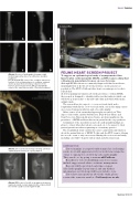
A
B
C
D
A
B
Above: A series of radiographs showing a right hind metatarsal fracture of a calf before and after treatment.
DP (A) and LM (B) views of the complete transverse fracture of the metatarsal prior to sedation, epidural and local anaesthetic blocking, reduction of the fracture and cast placement. DP (C) and LM (D) views of the same fracture after 12 weeks treatment.
Above: CT reconstruction image showing a femoral capital physeal fracture in a young alpaca.
Above: MRI scans of a sti e in an alpaca showing a ruptured cranial cruciate ligament (A), compared with the normal caudal cruciate ligament (B).
Issue: Summer
Summer 2016 11
