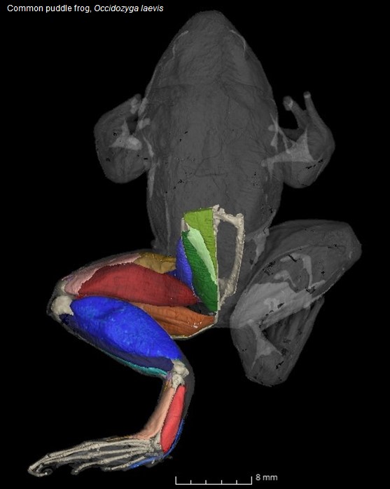
Hop to it: New research explores the muscle anatomy of frogs in relation to their movement
Researchers from the Royal Veterinary College (RVC) and UCL have uncovered anatomical differences between species of frogs specialising in different locomotor styles. Jumping, swimming, burrowing, walking and climbing frogs were all found to differ significantly in the size of their small hip and shank muscles. This provides new evidence of the muscles’ functional significance in a frog. Additionally, in the first analysis of its kind, the team found that evolutionary history determines the number of distinct muscles in the pelvis and thigh, while the separation of shank muscles is influenced more strongly by a frog’s movement.
The complex relationship between anatomy and function, and how it influences animal behaviour, has long posed a major challenge in evolutionary biology. Variation in muscle size and shape has historically been assumed to indicate differences in locomotor behaviour, where a larger muscle indicates higher functional importance, as more energy has been invested into its growth despite the associated physiological and anatomical costs.
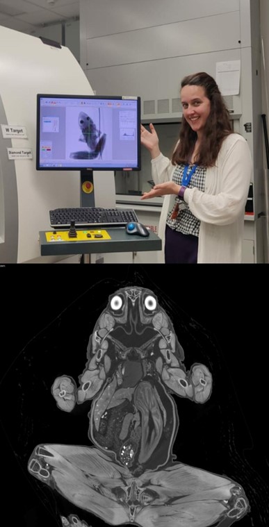
The team created the world’s largest dataset of digital dissections of any vertebrate group – including 30 species covering terrestrial, arboreal and aquatic habitats across Europe, Africa, North and South America and Oceania. Using specimens from museums, diffusible iodine contrast-enhanced microCT (diceCT) imaging was used to digitally dissect the muscle anatomy of each frog’s pelvis and hindlimbs.
Key findings showed that frogs specialising in jumping and swimming invested most heavily into shank musculature due to the strong requirements for powerful ankle extension. In contrast, burrowing frogs had the largest tarsal muscles, likely required to help them scoop surfaces with their feet.
The study presents important implications for future studies of frog palaeontology as the lengths of bones were found to not always be reliable predictors of muscle mass. 3D anatomical reconstructions are available to download for free to help teach amphibian musculoskeletal anatomy in dissection labs, provide veterinarians specialising in treating amphibians with greater insight, and offer educational resources for biological sciences being taught in schools and universities.
Dr Alice Leavey, first author who performed this study as part of her PhD thesis under the supervision of the RVC and UCL, said:
“While frogs have a highly conserved body plan, they use a highly diverse array of locomotor styles. Therefore, it was important for us to directly quantify and compare how the number of distinct muscles in sections of the body differ between species. This has important functional implications as it can impact the range of motion that body parts can perform.
“From schools and universities to scientists and vets, the most exciting part about this work is the massive potential for future research and education through the publication of the 3D reconstructions and annotations. We’ve even had requests from video-game developers to use them to help create more realistic characters. The list of potential uses is endless.”
Dr Christopher Richards, co-author of the paper, and Lecturer in Basic Sciences at the RVC said:
“This is an extremely exciting time to study anatomy. The digital imaging and analysis techniques that we used in our study allow us to resolve long-standing questions about anatomical structure-function relationships across an unprecedented range of taxa. Moreover, the digital reconstructions we have shared can continue to be used in teaching and research for years to come.”
Co-author, Dr Laura Porro, Associate Professor at UCL, said:
“Recent advances in new staining techniques, high-resolution imaging, and 3D visualisation have opened new possibilities for understanding human and animal anatomy to a level of detail that was previously not possible.”
The study “Comparative muscle anatomy of the anuran pelvis and hindlimb in relation to locomotor mode” was published in the Journal of Anatomy and was funded by the Biotechnology and Biological Sciences Research Council (BBSRC) via the London Interdisciplinary Doctoral training programme.
Notes to Editors
Reference
Leavey A, Richards C T., Porro L B. (2024) Comparative muscle anatomy of the anuran pelvis and hindlimb in relation to locomotor mode, Journal of Anatomy. https://doi.org/10.1111/joa.14122
For media enquiries, please contact:
- Jasmin De Vivo at jasmin.devivo@plmr.co.uk or rvc@plmr.co.uk
- Press Line: 0800 368 9520
About the RVC
- The Royal Veterinary College (RVC) is the UK's largest and longest established independent veterinary school and is a Member Institution of the University of London.
- It is one of the few veterinary schools in the world that hold accreditations from the RCVS in the UK (with reciprocal recognition from the AVBC for Australasia, the VCI for Ireland and the SAVC for South Africa), the EAEVE in the EU, and the AVMA in the USA and Canada.
- The RVC is ranked as the top veterinary school in the world in the QS World University Rankings by subject, 2024.
- The RVC offers undergraduate and postgraduate programmes in veterinary medicine, veterinary nursing and biological sciences.
- The RVC is a research-led institution, with 88% of its research rated as internationally excellent or world class in the Research Excellence Framework 2021.
- The RVC provides animal owners and the veterinary profession with access to expert veterinary care and advice through its teaching hospitals and first opinion practices in London and Hertfordshire.