New research by the RVC could hold key to new treatments for Cancer and Neurodegeneration
A new study, led by the RVC, has found a new interaction within cells that could lead to the development of novel therapies for pathological conditions in humans and animals such as cancer and neurodegeneration.
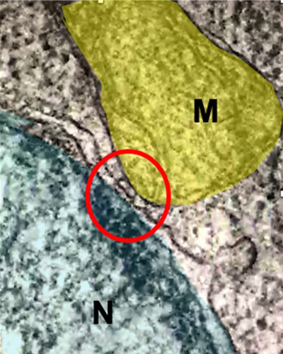
This discovery of direct interaction between mitochondria and nuclei through membrane contact sites expands the understanding of mitochondrion-to-nucleus communication and intracellular signalling.
In mammals, mitochondria are vital organelles acting as primary sources of energy. They are the most successful example of endosymbiosis, the process through which eukaryotic cells host a prokaryote with membrane bound nucleic acid, and play a part in several key processes spanning hormone synthesis and programmed cell death.
Despite the successful integration, they retain their own DNA, making the interplay with the nucleus - which contains the genomic DNA - a key aspect in the homeostasis of cells and tissues.
Until very recently, it was understood that mitochondria communicate to the nucleus alterations in the cellular environment to increase the transcription of genes involved in the preservation of mitochondrial and cellular function. This process, known as mitochondrial retrograde response (MRR), is ascribed to the deregulation of several pathologies including cancer and neurodegeneration.
The team, led by Professor Michelangelo Campanella, Chair in Pharmacology; Head of the Mitochondrial Cell Biology and Pharmacology Research Unit at the RVC, utilised breast cancer cells of different stages of aggressiveness isolated from human, dogs and cats. In these three species, the evolution of cancer of the mammary gland (e.g. breast cancer) meant that their susceptibility to chemotherapy was proven to be associated with the degree of contact sites between mitochondria and nuclei.
Various molecular pharmacology protocols were used throughout the study to induce and control the interaction between mitochondria and nuclei. Added to this, cutting edge techniques of fluorescent imaging enabled the team to map the dynamic nature of the interaction and transmission electron microscopy to picture the ultrastructure of Nucleus-Associated Mitochondria (NAM).
The team’s breakthrough demonstrates that MRR is mediated by specific points of contact within the NAM. The discovery of discrete anatomic compartments offers the possibility to target these selectively and regulate this inter-organelle communication, paving the way for new and improved strategies to impact diseases.
Professor Michelangelo Campanella, Chair in Pharmacology; Head of the Mitochondrial Cell Biology and Pharmacology Research Unit at the RVC, said:
“This study is the first of its kind to unveil the association between the mitochondrion and nucleus to be a regulated process and can be used to identify how this interplay can be pharmacologically controlled.
“The impact of this discovery is likely bigger than the advanced comprehension of mammalian cells physiology and pathology, embracing aspects of evolution. The co-existence of distinct DNAs is just partially understood and how genes from the mitochondria are transferred to genomic DNA is completely unknown. Our research group has now started investigating the molecular determinants of membrane tethering at NAM and the involvement of these inter-organellar communication in several disease models. This is truly ground-breaking as it will allow us to develop ways of correcting mitochondrial signalling in pathological conditions including cancer and neurodegeneration.”
This research was funded by the Biotechnology and Biological Sciences Research Council (BBSRC), the Petplan Charitable Trust and the European Research Council.
Research reference
This full paper is available at: https://advances.sciencemag.org/
Notes to Editors
For more information please contact:
- Jasmin De Vivo (Jasmin.DeVivo@plmr.co.uk or rvc@plmr.co.uk
- Press Line: 0800 368 9520
About the RVC
- The Royal Veterinary College (RVC) is the UK's largest and longest established independent veterinary school and is a Member Institution of the University of London. It was the first in the world to hold full accreditation from AVMA, EAEVE, RCVS and AVBC.
- The RVC is the top veterinary school in the UK and Europe, and ranked as the world’s second highest veterinary school in the QS World University Rankings by subject, 2020.
- The RVC offers undergraduate and postgraduate programmes in veterinary medicine, veterinary nursing and biological sciences.
- In 2017, the RVC received a Gold award from the Teaching Excellence Framework (TEF) – the highest rating a university can receive.
- A research led institution with 79% of its research rated as internationally excellent or world class in the Research Excellence Framework 2014.
- The RVC provides animal owners and the veterinary profession with access to expert veterinary care and advice through its teaching hospitals and first opinion practices in London and Hertfordshire.
You may also be interested in:
-
New collaboration between the RVC and UCB seeks to better understand diseases of excessive bone formation

Research, led by the RVC, will explore disease causing mechanisms of two severe bone diseases …