Alpaca Benefits from a Collaborative Approach After Trauma
Clinical Connections – Autumn 2024
David Bolt (Senior Lecturer in Equine Surgery) and Richard Meeson (Professor of Orthopaedics and Head of the Small Animal Orthopaedic Service) outline the case of an alpaca who benefited from the transdisciplinary approach of the RVC.

A two-year-old male Huacaya alpaca was treated at the RVC Equine Referral Hospital by the equine specialists, in collaboration with the RVC Small Animal Referrals’ Orthopaedics Service.
Dexter had sustained a traumatic right shoulder luxation approximately a week prior to referral. He presented with right forelimb lameness.
On presentation, Dexter was bright, alert and responsive. His general physical examination was normal, but he was lame on the right forelimb at walk. There was a reduced cranial phase to the stride.
Focal swelling present over the right shoulder region with mild focal pain on palpation was noted.
Diagnostic tests
A CT scan identified luxation of Dexter’s right shoulder joint with a large associated defect in the humeral head and flattening of the glenoid cavity of the scapula. There was dystrophic mineralisation and distension of the synovium and joint capsule, likely representing proliferative synovitis and possible haemarthrosis.
The scan also revealed complete displaced comminuted subacute and traumatic fracture of the acromion of the scapula. Additionally, there were mild changes to the proximal aspect of the bicipital tendon, suggestive of mild tendinopathy. This was considered to be a consequence of the luxation or normal variation.
Surgery
The surgery was a collaboration between the Equine and Small Animal Orthopaedic teams. Dexter was anaesthetised and positioned in left lateral recumbency. A brachial plexus block was performed by the RVC Anaesthesia and Analgesia team to provide additional analgesia.
A curved craniolateral incision was made, extending from the scapular neck to the mid-diaphysis of the right humerus. The joint capsule was incised after tenotomy of the infraspinatus tendon.
The humeral head was found to be craniolaterally luxated and was manipulated back into the glenoid cavity. Cortical screws and washers were then placed in the centre of the scapular neck and the proximal humerus behind the greater tubercle.
A tunnel was also created through the greater tubercle, from cranioventral to caudodorsal. Together they provided the anchorage for the artificial ligament placement – which included three different loops of a high-tension dual core artificial suture.
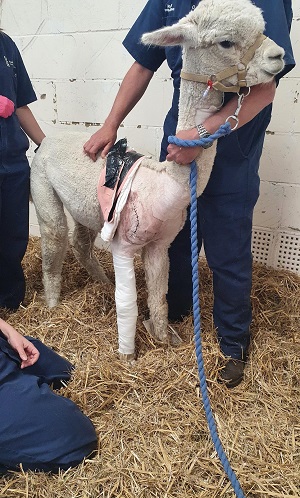
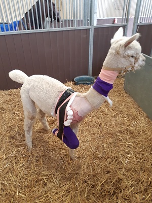
After the joint capsule and overlying soft tissues were closed, a dressing was placed on the limb, in addition to a Velpeau bandage.

Recovery and outcome
Dexter recovered uneventfully from surgery and anaesthesia. Radiographs were obtained after the procedure. The humeral head had remained in the correct position and all implants were intact.
The Velpeau sling and all skin sutures were removed three weeks after surgery and Dexter started weight-bearing and ambulating immediately. He was discharged from the hospital four weeks after the procedure.
Physiotherapy, consisting of passive flexion and extension of the right forelimb, was already initiated during Dexter’s hospitalisation and continued at home. After two months turn-out in a small paddock with a companion, Dexter was turned out with the remainder of the herd. His owners report that he has made a made a full recovery.
Dexter’s case illustrates the value and strength of teamwork and the use of pooled resources to provide care for all species of sick and injured animals at the RVC.
Shoulder reconstruction surgery is more commonly performed in RVC Small Animal Referrals and combining the expertise and experience of small animal surgery specialist Professor Meeson and large animal surgery specialist Dr Bolt resulted in a good outcome for Dexter and an enjoyable opportunity to share ideas and work together.
The surgeons were expertly supported by nursing teams from both the Equine Referral Hospital and the Queen Mother Hospital for Animals during the procedure. Anaesthesia in camelids can be very challenging and the RVC’s Anaesthesia and Analgesia Service facilitated a safe and successful procedure and also provided multimodal analgesia.
Dexter’s aftercare after surgery was provided by RVC BVetMed rotation students and training scholars (residents and interns) who all enjoyed being involved in this unusual case.
