A New Dimension in Visualisation
Clinical Connections – Autumn 2021
Maria-Christine Fischer, Lecturer in Ophthalmology
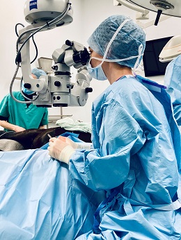
The new ophthalmic operating microscope Zeiss Lumera 700 was purchased with an Animal Care Trust (ACT) grant and has provided a vast amount of benefits, not only to our patients, but also to research, veterinary education, and specialist training.
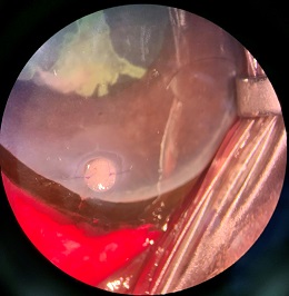
The operating microscope provides us with an exceptional surgical experience as its technology offers a greater depth and width in the field of view and the Stereo Coaxial Illumination technology allows us to visualise every anatomic detail with perfect clarity. This is a big advantage for routine procedures contributing to shorter anaesthetic times and has proven to be especially helpful when performing lens removal surgery in small exotic eyes.
Together with the new Alexos 3 Phaco system from An-Vision, also funded by the ACT, it is now possible to perform microincisional cataract surgery in even smaller exotic eyes.
Another advantageous feature of Zeiss Lumera 700 is the video screening. It has enabled an improved communication and interaction with the team during microsurgical procedures. The anaesthetists are now able to visualise the surgery and the response of the patient the specific drugs required for ophthalmic procedures. This is giving the team useful information on depth of anaesthesia, analgesia, and effect and duration of neuromuscular blocking agents that keep the globe in a central position for the procedure.
The nursing team can now anticipate the next move during the case and for example get the artificial intraocular lens or additional instruments ready. Further, our ophthalmology theatre nurses can now apply their knowledge when explaining surgical procedure to veterinary and nursing students, which has been very motivating for their professional development. Being able to watch the surgery live has enable everyone to get involved and to feel more ‘part of it’. This has led to a very enthusiastic and efficient collaboration of the team members and made challenging ophthalmic procedures something to look forward to. The video recording capacity enables the joint review and analysis of the procedures with the team aiming to refine surgical techniques.
Zeiss Lumera 700 is also equipped with an integrated keratometer. With this ring of light, we can visualise the corneal curvature in real time and it has proven a great help during corneal suture placement when performing cataract and corneal graft surgery. By monitoring the uniformity of the corneal sutures placed intraoperatively we can minimise postoperative corneal astigmatism and achieve better visual outcomes for our patients. A final year student, Monika Salkauskaite, has based her clinical research project on this topic. Under guidance of the ophthalmology team she successfully developed a protocol for its intraoperative use and for measurements that can objectively assess changes in the corneal curvature. This will find application in future scientific projects aiming to reduce visual deficits associated with corneal astigmatism.
Advancements in equine ophthalmic surgery
The presence of the Zeiss Lumera 700 at the Queen Mother Hospital for Animals (QMHA) allowed the former QMHA
Zeiss microscope to be moved to the Equine Referral Hospital permanently and facilitates immediate vision-saving intervention for our equine patients. This microscope, together with the new phacoemulsification system from An-vision, which is also equipped for cataract removal in horses, has increased the range of ophthalmic microsurgical procedures we can offer.